Glaucoma
In India, glaucoma is the leading cause of irreversible blindness with at least 12 million people affected and nearly 1.2 million people blind from the disease. More than 90 percent of cases of glaucoma remain undiagnosed in the community. Glaucoma prevalence increases with age. Often called the “sneak thief” of sight, people with most forms of glaucoma do not have symptoms until the optic nerve is already severely damaged. If diagnosed early, the disease can be controlled and permanent vision loss can be prevented.
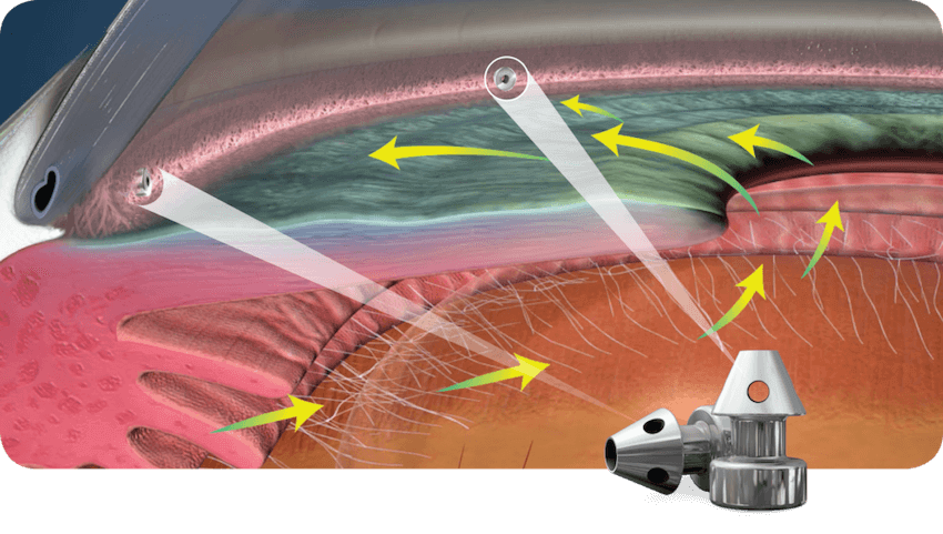
What causes glaucoma? Glaucoma occurs when the normal pressure inside the eyes (intraocular pressure or IOP) increases because the aqueous humor fluid – which usually flows in and out of the eye – is unable to drain. Over time, this fluid buildup damages the optic nerve, the structure that sends visual signals from your eyes to your brain. Underlying reasons for this usually relate to the type of glaucoma you have.
Symptoms of Glaucoma
In most cases, glaucoma is asymptomatic (has no symptoms). By the time an individual experiences decreased vision, the disease is frequently in its latter stages. Since early warning signs of glaucoma are rare, it is important especially for those at risk to have regular medical eye examinations every one or two years.
Patients with chronic glaucoma may not be aware of any symptoms because the disease develops slowly and they rarely notice loss of peripheral vision. Patients with an acute form of glaucoma (acute angle closure) may develop severe symptoms because ocular pressure rises quickly and they may experience:
- Blurred vision, especially at night
- Halos or rainbows around lights
- Severe headaches or eye pain
- Nausea
Common Types of Glaucoma
Acute Angle Closure GlaucomaAcute closure of the peripheral drainage angle, characterized by a sudden increase in intraocular pressure.
Primary Angle Closure GlaucomaThe iris obstructs the eye’s drainage angle in a slow, progressive fashion.
Primary Open Angle GlaucomaThe drainage angle is open but does not allow fluid to drain adequately for unknown reasons.
Pseudoexfoliation GlaucomaDeposits of a fibrillary material that may contribute to the obstruction of the fluid drainage from the eye.
Pseudoexfoliation GlaucomaDeposits of a fibrillary material that may contribute to the obstruction of the fluid drainage from the eye.
Pigmentary GlaucomaPigment dislodged from the iris obstructs the eye’s drainage structures.
Angle Recession GlaucomaScar tissue from previous trauma obstructs the outflow of fluid.
Neovascular GlaucomaVarious disorders cause blood vessels to proliferate on the iris and in the eye’s drainage structures.
Normal Tension GlaucomaGlaucoma that develops despite eye pressure in the normal range.
Childhood Glaucoma or Pediatric GlaucomaChildhood glaucoma, also referred to as congenital glaucoma, pediatric glaucoma or primary infantile glaucoma occurs in babies and young children.
Glaucoma Risk Factors
Although glaucoma is most common in adults over the age of 40, susceptibility is not determined by age alone. A genetic predisposition of those with a family history of the disease , are at a particularly increased risk. Studies have shown individuals at greater risk for glaucoma may fit one or more of the following criteria
- Are over the age of 60
- Have a family history of the disease, elevated intraocular pressure
- Have diabetes or hypertension
- Are very nearsighted or farsighted
- Steroid Users
- Have had an eye injury
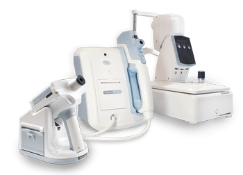
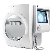
Expert Diagnosis for Early Detection
To achieve an accurate assessment, experienced ophthalmologists perform a comprehensive glaucoma screening that consists of non-invasive, pain-free procedures:
TonometryThis test measures the pressure inside your eye.
Visual FieldThis test checks for vision loss in your side or peripheral vision.
Spectral Domain OCTNewer diagnostic studies using computer-imaging technology such as spectral domain optical coherence tomography (OCT), now permit precise measurements of the retinal nerve fiber layer that cannot be visualized by the unaided human eye. This test helps monitor and detect optic nerve loss over time.
Optic Disc PhotographyOptic nerve photographs document the severity of damage to the nerve and are used to monitor changes over time.
PachymetryBecause corneal thickness can influence your eye pressure reading, this test measures the thickness of your cornea.
GonioscopyThis exam looks at the drainage angle in your eye.
Treatments
Eye DropsCertain prescription eye drops decrease intraocular pressure by reducing the amount of fluid your eye produces. Several different classes of glaucoma medications are available to provide pressure reduction including beta-blockers, prostaglandin analogues, alpha-adrenergic agonists, miotics, Rho kinase inhibitors, and oral and topical carbonic anhydrase inhibitors. These medications work by either reducing the rate at which fluid in the eye is produced or by increasing the outflow of fluid from the eye.
Oral MedicationsLike eye drops, oral prescription medications help reduce pressure inside your eye.
Laser TherapyUsing a laser beam, your ophthalmologist opens clogged channels inside the eye, releasing fluid build-up. Laser therapy is an outpatient procedure.
Filtering SurgeryFiltering surgery to create a new passage for fluid drainage. Surgery is usually reserved for cases that cannot be controlled by medication and after appropriate laser treatment.
Implant SurgeryTo enhance filtering surgery, your ophthalmologist may insert tiny drainage devices or “aqueous shunts” to keep the surgically created drainage opening from closing.
MIGSMinimally invasive glaucoma surgery (MIGS) has been developed in recent years to lower eye pressure and prevent progression of glaucoma. MIGS procedures are indicated in certain types of glaucoma and work by using microscopic-sized equipment and tiny incisions.
Timings
Mon - Sat : 9:30 AM - 4:00 PM
Sunday : 9:30 AM - 1:00 PM
Why Choose Eswar Eye Hospital?
- Serving the Locality - When you choose Eswar Eye Hospital, you choose oldest eye care provider serving this locality with advanced treatments and high-quality patient care.
- Patient-Centered Care and Support - This chronic disease requires ongoing medical care and management. In the process of caring for you, our eye care professionals get to know you as a person, not just a patient. They provide the expertise, education and support you need to manage your condition.
- If You Have Any Questions Call Us On (+91) 9177451477
- Make an Appointment